Posts

Showing posts with the label Inverted Fluorescence Microscope

New D-LEDI from Nikon (update - 2022-08-30)

Image
- Due to the mercury lamp box spare part will be no longer supplied, thus the company recommend to replace our current light source with the LED. - LED box contains 4 LED lights, 1) 385 nm, 2) 475 nm, 3) 550 nm, 4) 621 nm as shown in the picture below. - Showing the excitation and emission profiles with current available filters that we have ( https://minicore-rarc.blogspot.com/2018/05/upright-fluorescence-microscope.html ) If you would like to try it by yourself -- I would recommend to visit:  https://www.bdbiosciences.com/en-nz/resources/bd-spectrum-viewer - Testing set that we would like to try with the new LED light source - Set 1      - Nucleus – Dapi (LED 385 – filter UV-2A)      - Rad51 - Alexa fluor 555 (LED 550 – filter TexasRed)       - H2Ax -- Alexa fluor 488 (LED 475 – filter FITC) - Set 2      - Nucleus - Dapi (LED 385 – filter UV-2A)      - Rad51 - Alexa fluor...
Current status of optic fiber condition (switch from inverted microscope to upright)

Image
  As of 21 Aug 2021, one dim spot was observed. Please take good care of the fiber optic.

Update on the microscope condition

Image
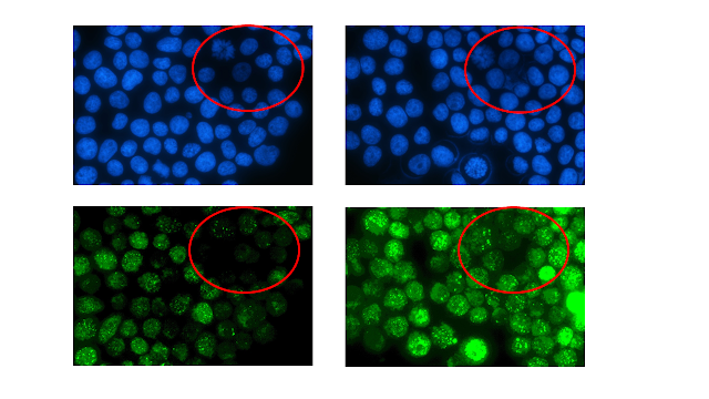
We have just learnt from the Nikon technician that our fiber optic has been damaged. This causes a shadow background while visualizing the sample with a mercury lamp (attached herewith), not a bright field. Besides, all the lenses have been contaminated by the immersion oil. This means that there might be some spilled over while rotating the objective lens.   Please make sure to clean "all the objective lenses" properly after finishing your work.  Learn from my deposited video if you are not sure how to properly clean.  The 60X objective lens of the upright microscope does not work properly anymore and I decided not to replace it with a new one since it is extremely expensive.  I decided to switch the optic fiber between inverted and upright. Thus, the shadow will instead appear in the inverted microscope in the cell culture-micro room. We need to place an order for the new optic fiber and I do not know when it will be approved (cost perhaps  >...

ZEISS ZEN lite Free Imaging Software for Microscopy (update: 2025-05-21)

Image
I am not a custodian of this scientific instrument but I have to use it. I asked the custodian to make video in order for the users to self-learn and remind. To download the software; https://www.zeiss.com/microscopy/en/products/software/zeiss-zen-lite.html Manual The imageJ can open the .czi file. How to use ZEN lite (require dongle to analyze the full mode) Location: cell culture II Booking: Log-book

Inverted Fluorescence Microscope (update: 2025-10-22)

Image
Filter cubes Up to 5 slots, one is left in blank for the bright field Available filters in our facility; 1. UV2A (Longpass filter) 2. B2A (Longpass filter) 3. G2A (Longpass filter) 4. Texas red (Bandpass filter) 5. FITC (Bandpass filter) 6. DAPI (Bandpass filter) -- Phase contrast -- Lens: Plan Fluor 4X/0.13NA Ph1 Plan Fluor 10X/0.3NA Ph1 S Plan Fluor 20X/0.45NA (adjustable correction collar) Ph2 S Plan Fluor 40X/0.60NA (adjustable correction collar) Ph2 S Plan Apo 60X/0.7NA (adjustable correction collar) Only trained personnel will be allowed to change the filters by themselves. Check out type of filters:  https://bityl.co/C3BV Manual Nikon Ti-U (Eng-full manual) Fluorescence microscope (Ti Series) - SOP from Tassanee Lerksuthirat Link to download NIS software (only viewer - free) https://www.microscope.healthcare.nikon.com/en_AOM/products/software/nis-elements/viewer Instructional video Location Cell culture room, microbiology area Only person who has ...